Article
How to use an ultramicrotome for challenging sample preparation
Some samples refuse to cooperate. Soft polymers compress, biological tissues distort, and brittle ceramics fracture the moment the knife makes contact. At the ultrathin scale required for electron microscopy, even the smallest imperfection can render a section unusable.
This is where ultramicrotomes come in. These precision instruments use diamond or glass knives to cut sections as thin as 50 to 100 nanometers. While ultramicrotomes make nanoscale sectioning possible, handling difficult materials such as elastomers, metals, and multi-layered composites demand more than placing a sample in the machine. To get clean, reproducible sections, users must fine-tune cutting conditions, control environmental factors, and adapt the sample preparation method for each material.
Understanding the Ultramicrotome
An ultramicrotome is a highly specialised instrument designed to cut ultrathin sections for transmission electron microscopy (TEM) and other high-resolution imaging techniques. Unlike standard microtomes, which cut to micrometre thicknesses, ultramicrotomes achieve nanometre precision using an incredibly sharp knife alongside an ultra-fine feed system.
Cutting involves several key steps:
- Sample Mounting: The specimen, embedded in resin or frozen for cryo-ultramicrotomy, is secured in a specimen holder.
- Knife Engagement: The cutting edge moves steadily against the sample, generating ultrathin slices.
- Section Collection: The section floats on a liquid surface, usually water, to prevent damage before being transferred onto a TEM grid or another substrate.
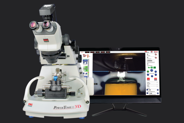
While ultramicrotomes provide the mechanical precision needed for sectioning, achieving high-quality sections, especially from difficult materials, depends on proper technique. Systems like RMC Boeckeler’s PowerTome are engineered for nanoscale precision, combining robust power-driven mechanics with ergonomic control for advanced sample sectioning.
Utilising an Ultramicrotome for Challenging Sample Preparation
Challenging samples cannot be prepared using a one-size-fits-all approach. Each material presents unique obstacles that require precise adjustments to sectioning techniques. The following factors are key to achieving high-quality sections from difficult specimens.
1. Optimising Cutting Parameters
Fine-tuning the ultramicrotome’s settings is the first step toward achieving clean, reproducible sections.
- Cutting Speed: A slow, controlled stroke between 0.1 and 0.5 mm per second minimises tearing and distortion.
- Knife Angle: Adjusting between 45 and 55 degrees lowers compression in soft samples and reduces chipping in brittle ones.
- Feed Rate: Lowering the feed rate applies less force, reducing the likelihood of fractures in fragile materials.
2. Choosing the Right Knife for the Material
Selecting the appropriate ultramicrotome knife ensures a clean cut without damaging the sample.
- Diamond ultramicrotome knives offer the highest precision and durability, making them ideal for brittle materials like ceramics, metals, and nanocomposites.
- Glass knives work well for biological and polymer samples but dull quickly and require frequent replacement.
- Cryo-knives prevent deformation in temperature-sensitive samples, stabilising soft or hydrated materials for clean sectioning. PowerTome ultramicrotomes from RMC Boeckeler support all major knife types, making them adaptable for a wide range of materials.
Regularly rotating or replacing the ultramicrotome knife ensures consistent performance and prevents unnecessary resistance that can lead to artefacts.
3. Adjusting for Material-Specific Challenges
Each type of sample presents unique difficulties that require specialised solutions.
- Soft polymers and biological tissues tend to compress or stretch under the knife. Cryo-ultramicrotomy stabilises them by freezing the material, preventing deformation.
- Brittle materials, such as metals, ceramics, and hard polymers, crack easily. A diamond knife, lower cutting force, and resin impregnation help prevent fractures.
- Multi-layered composites, including thin films, coatings, and laminates, can separate mid-sectioning. Adjusting the knife angle and ensuring even cutting pressure helps maintain structural integrity.
4. Using Cryo-Ultramicrotomy for Soft or Hydrated Samples
Standard sectioning methods struggle with materials that distort or smear under normal conditions. Cryo-ultramicrotomy stabilises samples by freezing them before cutting. This technique is particularly useful for:
- Hydrated tissues that collapse at room temperature.
- Rubbers and elastomers that compress or stretch when sectioned.
- Lipid-based samples that smear rather than forming clean sections.
By freezing the sample, cryo-ultramicrotomy allows for sharper, more consistent ultrathin sections.
5. Ensuring Proper Section Collection
Even a perfectly cut section can be ruined if mishandled during collection.
- Floating Sections on Water: Most ultramicrotomes feature a knife trough where sections float, reducing mechanical stress and preventing curling.
- TEM Grid Transfer: For electron microscopy, ultrathin sections must be lifted onto copper or nickel grids using specialised tools to avoid distortion.
- Automated Collection for High-Volume Work: In studies requiring thousands of sections, automated tape-collecting ultramicrotomes streamline the process and ensure consistency. Systems like the PT3D PowerTome enable precise serial sectioning with reliable automation features.
Proper handling makes sure sections remain intact for imaging and analysis.
Overcoming Common Ultramicrotomy Issues
These are the best ways to address common issues:
- Compression Artifacts: Sections appear squashed when the ultramicrotome knife applies too much pressure. Reducing the speed, adjusting the knife angle, or switching to cryo-sectioning can help.
- Chatter Marks: Striations on sections suggest vibration or uneven resin hardness. Providing stable mounting and using a fresh ultramicrotome knife often improves results.
- Tearing and Cracking: Brittle samples break under stress. A diamond ultramicrotome knife, lower feed rate, and resin support minimise damage.
Using advanced models like the PTPCZ PowerTome, which integrates high-definition video display, operators can observe sectioning in real time, reducing errors and improving consistency.
RMC Boeckeler and CN Tech: Ultramicrotomy Tools for Advanced Research
RMC Boeckeler, a division of Boeckeler Instruments, manufactures precision equipment for nanoscale research across life and materials sciences. Through a strategic partnership with CN Tech, these tools are now available with expert local support, making advanced ultramicrotomy more accessible to researchers.
Available from CN Tech, the PowerTome series forms the core of RMC Boeckeler’s ultramicrotomy range.
- The PTXL model offers extended X and Y axis travel for accurate sectioning of large or irregular specimens.
- The PTPCZ integrates high-definition video with real-time display, ideal for observation, training, and documentation.
- The PT3D supports a full 1 mm specimen advance and Visutrac™ cutting zone control, designed for 3D reconstruction and array tomography workflows.
With a modular, upgradeable design, each model offers the flexibility to support both current and future applications. This adaptability, combined with our local support, gives laboratories reliable access to RMC Boeckeler’s trusted ultramicrotomy systems.
The Right Tools for the Most Difficult Samples
Ultramicrotomy is a skill requiring practice, patience, and the right equipment. By optimising cutting parameters, selecting the appropriate ultramicrotome knife, and making material-specific adjustments, researchers can successfully section the most challenging of samples.
For laboratories that need precision and reliability, we provides a range of ultramicrotomy solutions, including the PowerTome Ultramicrotome, LN Ultra Cryo Attachment, and ATUMtome for automated high-volume sample collection. These tools ensure clean, reproducible sections for electron microscopy and other nanoscale imaging techniques.
With the right approach and ultramicrotome equipment, even the most uncooperative samples can be prepared successfully for high-resolution imaging.